Ultrasound is a simple, safe, painless diagnostic procedure that bounces high frequency sound waves off parts of the body and captures the returning echoes as images. There is no injection or radiation exposure associated with ultrasound.
Ultrasound is able to capture moving images of pelvic and abdominal function (including gallstones), breast abnormalities, the male reproductive system, the kidney and thyroid systems, as well as the developing fetus among other applications. When enhanced with a special Doppler technique, ultrasound can also capture moving blood images of large blood vessels and moving images of the heart using echocardiography.
Preparation for your ultrasound will depend on the type of exam; a representative will call you prior to your appointment to provide specific instructions, and review health and insurance information. Please bring previous imaging study results (x-ray, MRI, CT, etc.) such as reports, films or CD-roms, if available. Please arrive 15 minutes early to verify your registration.
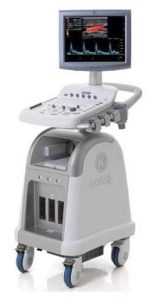
During the exam, you will lie on a cushioned table and gel will be applied to your skin; the gel acts as a conductor. A transducer, the hand-held device that sends and receives ultrasound signals, is moved over the area of your body being imaged. Images are instantly captured on a television-like monitor and transferred to film or videotape for a radiologist to review and interpret. Depending on the type of exam, it can take anywhere from 20-60 minutes.
After the exam, a radiologist who specializes in a specific area of the body will review your images (i.e., a body radiologist will review an ultrasound of your abdomen). The radiologist prepares a diagnostic report to share with your doctor. Your doctor will consider this information in context of your overall care, and talk with you about the results.
Ultrasound is able to capture moving images of pelvic and abdominal function (including gallstones), breast abnormalities, the male reproductive system, the kidney and thyroid systems, as well as the developing fetus among other applications. When enhanced with a special Doppler technique, ultrasound can also capture moving blood images of large blood vessels and moving images of the heart using echocardiography.
Preparation for your ultrasound will depend on the type of exam; a representative will call you prior to your appointment to provide specific instructions, and review health and insurance information. Please bring previous imaging study results (x-ray, MRI, CT, etc.) such as reports, films or CD-roms, if available. Please arrive 15 minutes early to verify your registration.
During the exam, you will lie on a cushioned table and gel will be applied to your skin; the gel acts as a conductor. A transducer, the hand-held device that sends and receives ultrasound signals, is moved over the area of your body being imaged. Images are instantly captured on a television-like monitor and transferred to film or videotape for a radiologist to review and interpret. Depending on the type of exam, it can take anywhere from 20-60 minutes.
After the exam, a radiologist who specializes in a specific area of the body will review your images (i.e., a body radiologist will review an ultrasound of your abdomen). The radiologist prepares a diagnostic report to share with your doctor. Your doctor will consider this information in context of your overall care, and talk with you about the results.